Dr. Aruna Seneviratne, MD. Mount Sinai Orthopedic Faculty Practice

Dr. Aruna Seneviratne, MD.
Mount Sinai Orthopedic Faculty Practice

mount sinai

877 636 7846

Book via ZocDoc Manhattan
Brooklyn, NY

Meniscus Tears

Updated 02/21/2015
Written and edited by Aruna Seneviratne, M.D

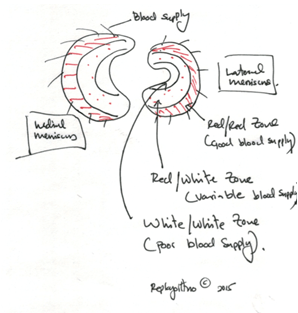
The meniscus is a C shaped piece of cartilage that resides between the two bones of the knee joint – the femur and the tibia. There are two menisci in each knee – one on the in-board side of the knee (medial meniscus), and one on the outboard side of the knee (lateral meniscus). They serve an important role in the function of the knee providing shock absorption, distribution of pressure, joint lubrication amongst other functions such as a secondary stabilizer of the knee.

Symptoms

Meniscus tears cause pain in the knee. Frequently there is swelling and clicking or a sensation of catching. Squatting and kneeling become painful and difficult. Participation in sports can be difficult. The pain can be quite severe at times.

Biology: Why does it occur?

Menisci can tear due to twisting or pivoting of the knee during sports activities, or other mechanisms. Sometimes the injury mechanism is so subtle that patients may not remember an event that caused their knee to hurt. The blood supply to the meniscus comes from the periphery of the meniscus where it is attached to the knee capsule, and penetrates the meniscus traversing centrally. As we age, the blood supply recedes and that has implications on how meniscus tears are treated.
meniscus

Common Diagnostic Techniques

History: Diagnosing the problem begins with a detailed history that your surgeon will obtain from you.
Physical Exam: A thorough physical examination is conducted by your surgeon.
X-Rays: Plain radiographs (X-Rays) are the most important diagnostic study that is performed initially. For the knee your surgeon will obtain specialized views. Usually these are weight bearing x-rays – i.e.: you will be standing for the x-rays.
MRI: Your surgeon may obtain additional studies such as an MRI to look more closely at the meniscus and other soft tissues.

Treatment

Treatment depends on a variety of factors including patient age, activity level, tear size, and location of tear. Older patients are usually treated non-operatively with physical therapy. If the tear is greater than 2cm in size the prognosis for becoming symptom free from non-surgical care is poor. If the tear is in an area of good blood supply, the tear may heal without surgery. Dr. Seneviratne will carefully evaluate each patient and recommend the optimum course of treatment factoring in all that is relevant to that patient. The goal is to treat patients with the least invasive manner.

Meniscus Surgery

The most common surgical treatment of meniscus tears is arthroscopic partial meniscectomy where a tiny camera is introduced into the knee via a tiny incision (keyhole surgery), and using arthroscopic instruments via a second keyhole, the torn piece of the meniscus is removed. The surgery takes about 20 minutes to complete and is performed under general anesthesia. This operation is one of the most common orthopedic operations performed in the United States.
Since the meniscus plays an important functional role in the knee, all attempts are made to preserve the meniscus and repair the tear if it is amenable to repair. Patient selection is important to ensure outstanding outcomes. Tears located in the vascular zone in younger patients are usually repaired. Dr. Seneviratne performs all three described techniques of repair – all inside arthroscopic repair, outside-in repair, and inside-out repair. Dr. Seneviratne has published techniques on meniscus repair.
Dr. Seneviratne will evaluate each patient individually and recommend a customized plan of surgical care.

After Surgery

Patients are allowed full weight bearing and are discharged from the surgical facility the same day. A cane maybe needed for a few days. Crutches are rarely needed. Most patients do not require narcotic pain medicines and can manage the pain with Tylenol or a NSAID such as Advil or Aleve. The knee will progressively start to feel better over the next few days to weeks. Dr. Seneviratne will see the patient back in the office for suture removal and to start physical therapy at about 7-10 days after the operation. Patients will need PT for about 6-8weeks. Running is possible in 6 weeks.

FAQ's

Infection, bleeding, damage to nerves and blood vessels, blood clots that form in your legs (DVT), blood clots that can break off and travel to your lungs causing a pulmonary embolus (PE). Knee stiffness is an additional risks. These risks are quite uncommon – less than 1% possibility. Possibility of requiring additional surgery does exist if the meniscus re-tears or if you have persistent pain for a variety of reasons.
Full recovery can be expected in about 3 months depending on the patient. Meniscus repairs take longer to heal.